
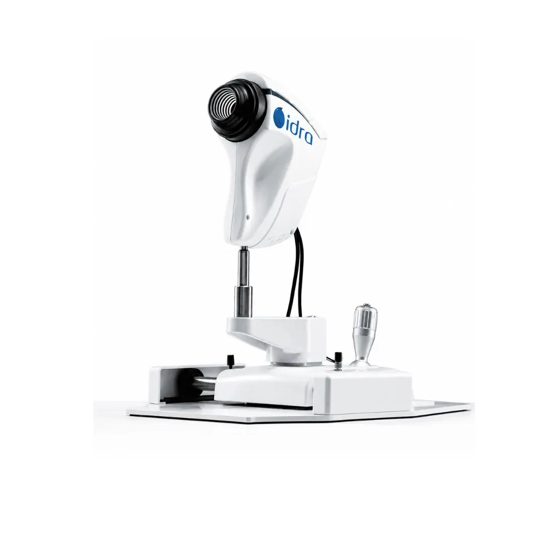
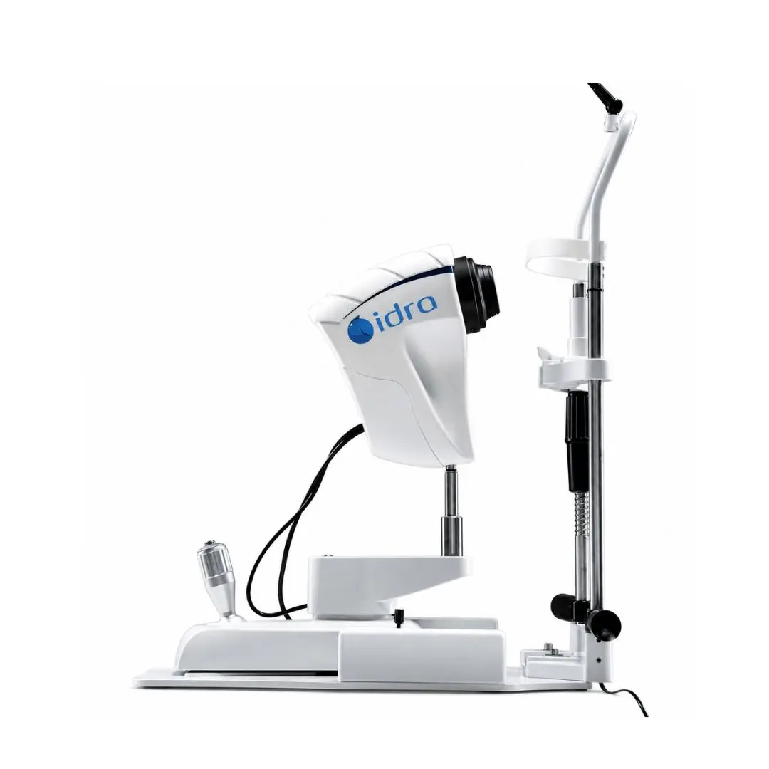
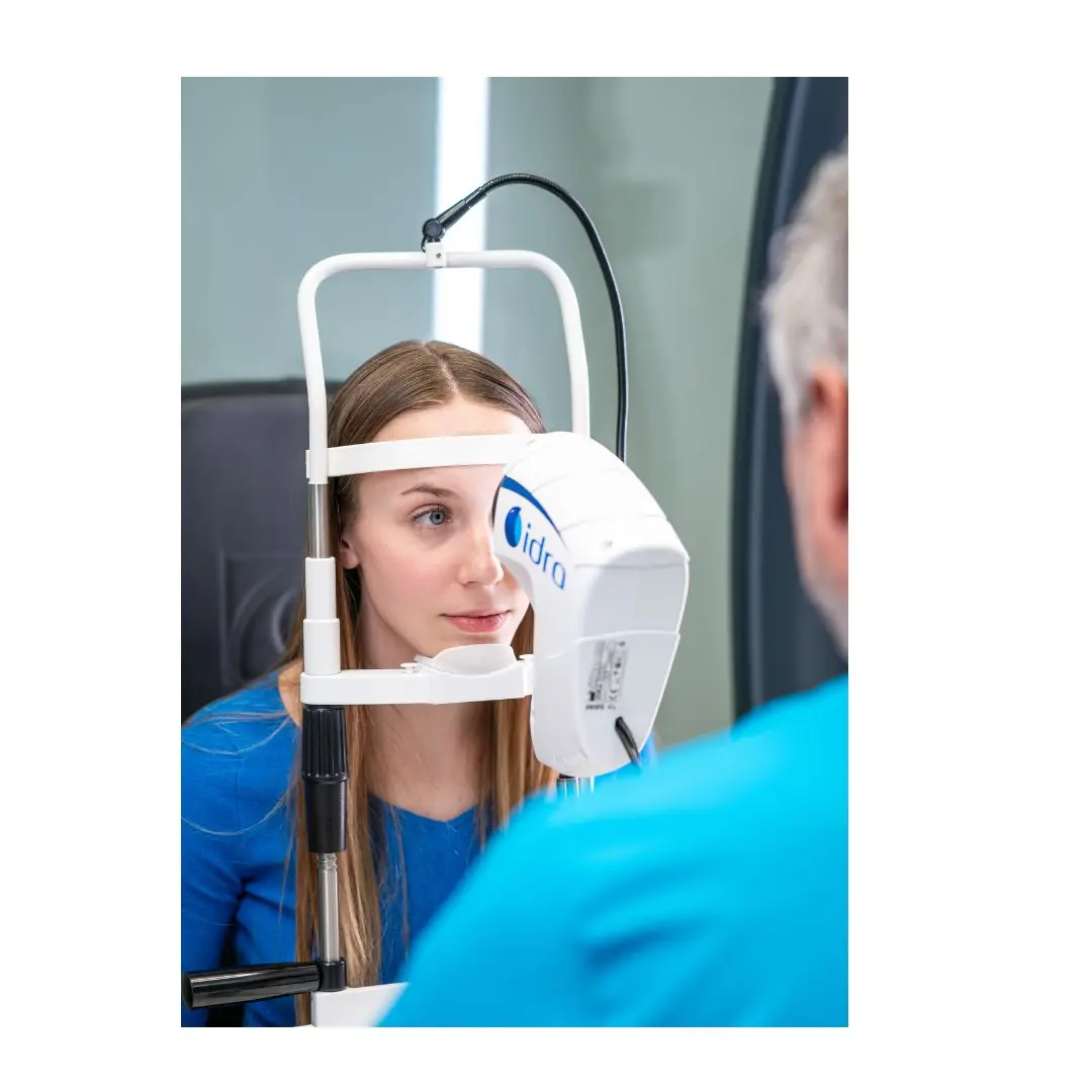
SBM Sistemi IDRA Dry Eye Diagnostic Platform
Evaluate tear-film stability, meibomian glands, lipid layer and ocular surface with the SBM Sistemi IDRA dry-eye diagnostic platform
The SBM Sistemi IDRA is a comprehensive ocular-surface diagnostic platform developed for rapid, detailed and noninvasive assessment of tear-film composition.
Its multimodal camera combines color imaging, near-infrared sensitivity and yellow-filtered acquisition with infrared, blue, red and white LED illumination. Two imaging cones—the main cone and Placido cone—support the different examinations available within the platform.
Depending on the selected software configuration, IDRA can evaluate tear-film lipid patterns, tear-meniscus height, noninvasive tear-breakup time, meibomian glands, blink quality, eyelid margins, bulbar redness, pupil diameter and white-to-white corneal diameter.
Automatic tools assist with NIBUT measurement, tear-film stability mapping, tear-meniscus measurement and, in the Full configuration, lipid-layer interferometry. The system also captures images and videos that facilitate documentation, patient education and comparison during follow-up.
IDRA is available in Basic, Plus and Full configurations. The exact examinations and automation features included depend on the purchased license.
Key Features
- Comprehensive dry-eye and ocular-surface assessment
- Noninvasive tear-film examination
- 8-megapixel imaging
- Color and near-infrared camera
- Automatic and manual focusing
- Multishot image and video acquisition
- Variable ISO control
- Infrared, blue, red and white LED illumination
- Main and Placido imaging cones
- Automatic NIBUT analysis
- Tear-film stability graph and breakup map*
- Automatic tear-meniscus measurement*
- Lipid-layer interferometry
- Meibography and gland-loss evaluation
- 3D meibography*
- Blink-quality analysis*
- Blepharitis and eyelid-margin imaging
- Bulbar-redness classification
- Pupillometry and white-to-white measurement
- Anterior-segment imaging
- Customizable examination and treatment-suggestion protocols
*Availability depends on the selected configuration.
Available Examinations
- Interferometry: Evaluates the interference pattern and characteristics of the tear-film lipid layer.
- Automatic interferometry: Automated lipid-layer assessment in the Full configuration.
- NIBUT: Noninvasive measurement of tear-film breakup time.
- NIBUT map: Displays the location and progression of tear-film breakup.
- Tear meniscus: Assesses tear volume through meniscus-height measurement.
- Meibography: Infrared visualization of the meibomian glands.
- 3D meibography: Three-dimensional representation of gland morphology and loss.
- Blink analysis: Evaluates blink patterns and incomplete blinks.
- Blepharitis examination: Documents eyelid margins and related structures.
- Bulbar redness: Compares conjunctival images with clinical grading scales.
- Pupillometry: Measures pupil diameter.
- White-to-white: Measures horizontal corneal diameter from limbus to limbus.
- Anterior-segment imaging: Captures clinical images of the ocular surface.
Configuration Comparison
| Examination | Basic | Plus | Full |
|---|---|---|---|
| Interferometry | Manual | Manual | Automatic |
| Tear meniscus | Manual | Automatic | Automatic |
| Automatic NIBUT | Yes | Yes | Yes |
| NIBUT map and stability graph | — | Yes | Yes |
| Meibography | Yes | Yes | Yes |
| 3D meibography | — | Yes | Yes |
| Blink-quality analysis | — | Yes | Yes |
| Blepharitis examination | Yes | Yes | Yes |
| Ocular-redness classification | Yes | Yes | Yes |
| Pupillometry | Yes | Yes | Yes |
| White-to-white measurement | Yes | Yes | Yes |
| Anterior-segment imaging | Yes | Yes | Yes |
| SKU | SBM-IDRA |
|---|---|
| Brand | SBMSistemi |
| Category | Dry Eye Analyzers |