
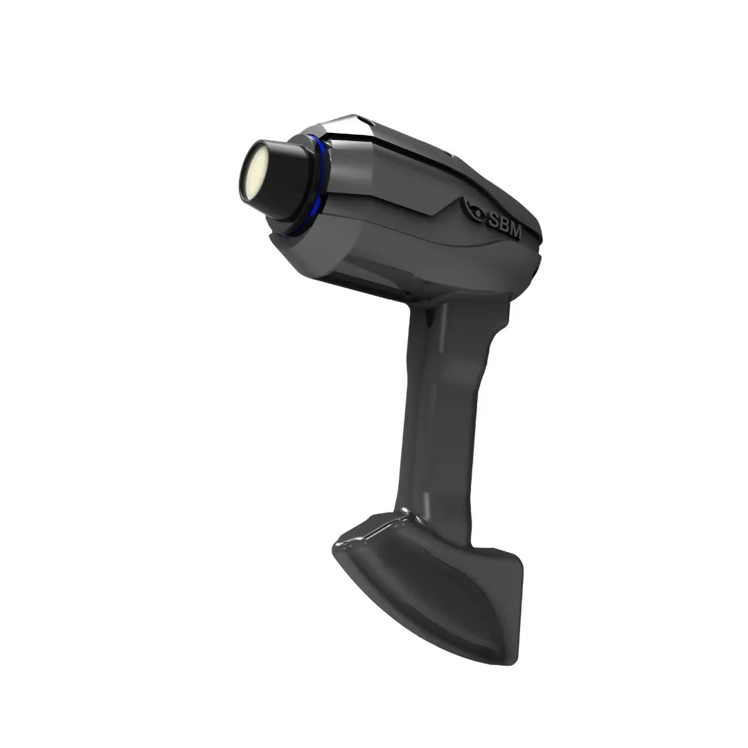
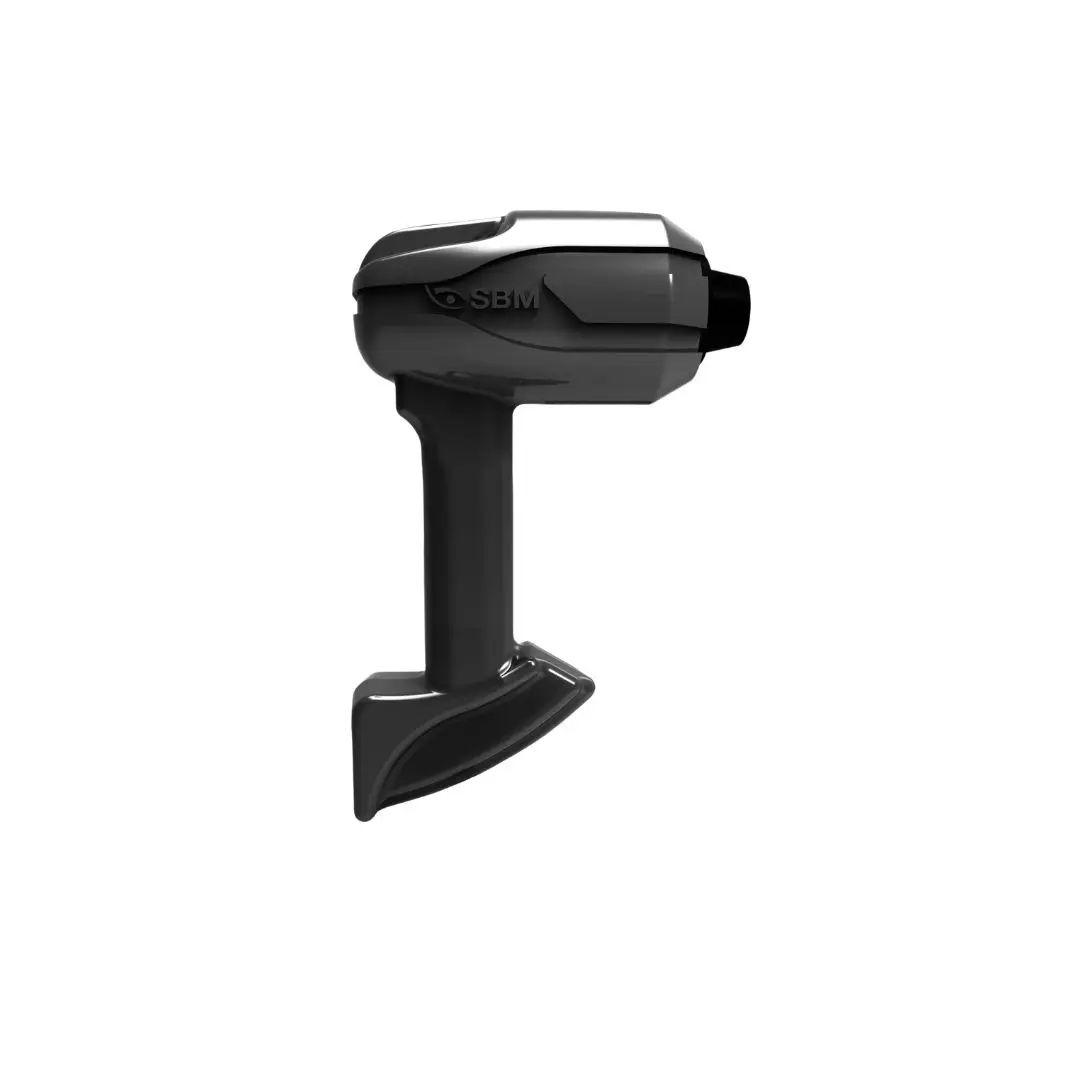
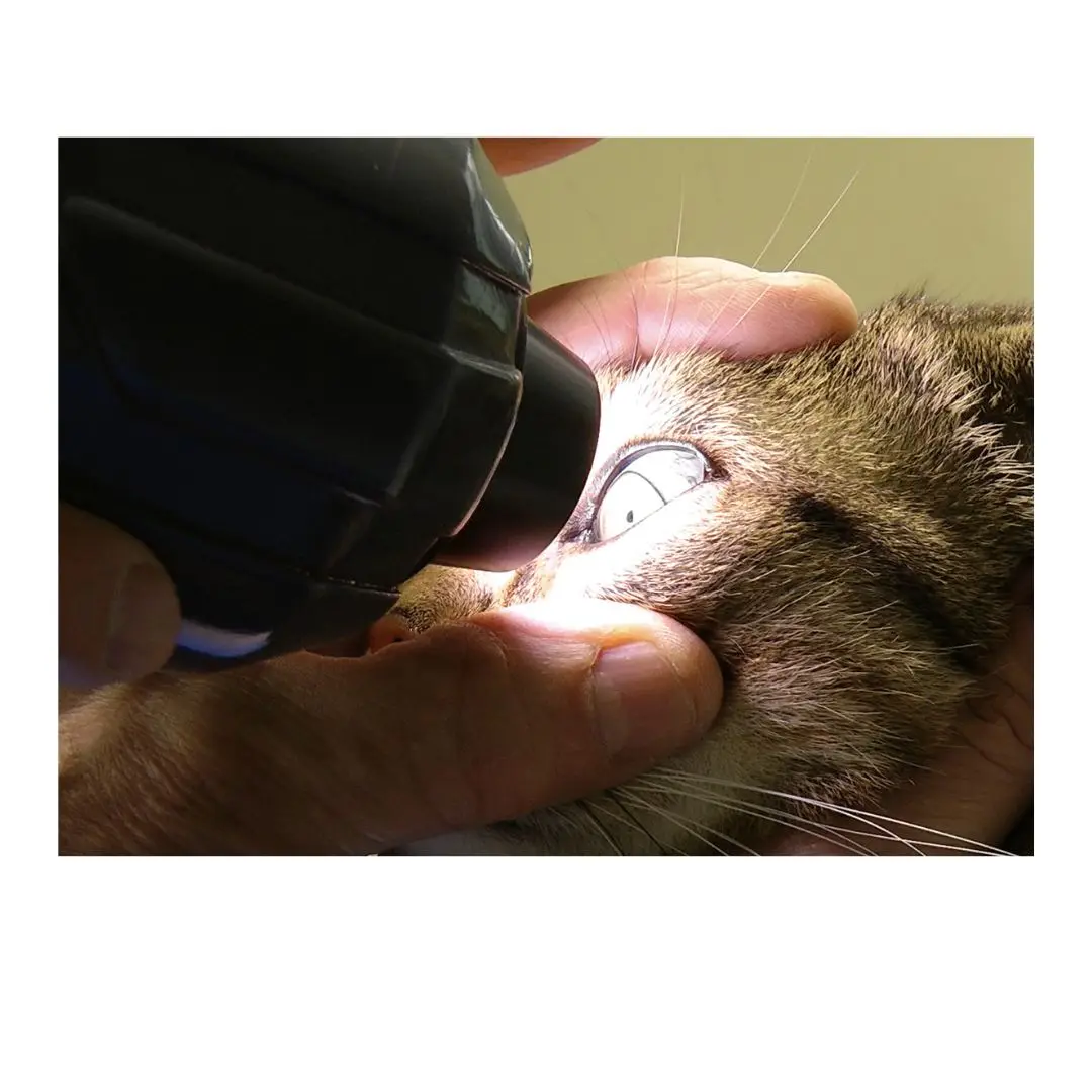
SBM Sistemi OSA-VET Veterinary Ocular Surface Analyzer
Evaluate animal tear film, meibomian glands and dry-eye parameters with the portable SBM OSA-VET veterinary ocular surface analyzer.
The SBM Sistemi OSA-VET is an integrated veterinary diagnostic platform designed to document and analyze the ocular surface and precorneal tear film in animals.
Its multimodal imaging system combines a 5 MP color camera with near-infrared sensitivity, a yellow filter, variable ISO control and multiple LED illumination sources. Dedicated interferometry and Placido cones support different examination techniques without direct contact with the ocular surface.
OSA-VET provides tools for evaluating tear-film quality and stability, including lipid-layer interferometry and non-invasive tear break-up time. It also supports infrared meibography, tear-meniscus assessment and white-to-white corneal diameter measurement.
The system can capture multiple images and video, facilitating documentation, comparison and longitudinal follow-up. Its portable configuration and compatibility with Windows-based systems make it suitable for veterinary ophthalmology practices, specialty hospitals, universities and mobile clinical environments.
Results must be interpreted by a qualified veterinary professional and considered alongside the animal’s clinical examination and other applicable diagnostic tests.
Key Features
- Veterinary ocular surface and dry-eye assessment platform
- Portable, integrated configuration
- Non-contact examination methods
- 5 MP image resolution
- Color camera sensitive to near-infrared light
- Integrated yellow filter
- Automatic and manual focusing
- Multishot image and video acquisition
- Variable ISO control
- Interferometry cone
- Placido cone
- Infrared, blue, red and white LED illumination
- Tear-film interferometry
- Non-invasive tear break-up time assessment
- Infrared meibography
- Tear-meniscus evaluation
- White-to-white corneal diameter measurement
- Image documentation and follow-up tools
- Compatible with Windows-based computer systems
- Suitable for veterinary clinical workflows
Examination Modules
Tear-Film Interferometry
Supports observation and documentation of the lipid component of the precorneal tear film using reflected-light patterns.
Veterinary Meibography
Captures near-infrared images of the meibomian glands and provides tools for image enhancement, gland-loss classification, gland-size measurement and follow-up comparison.
Non-Invasive Break-Up Time
Evaluates tear-film stability without applying fluorescein. Placido-ring reflections help identify changes and irregularities in the tear film.
Tear-Meniscus Assessment
Documents the tear meniscus along the eyelid margin to support evaluation of tear volume, height, shape and regularity.
White-to-White Measurement
Measures the horizontal corneal diameter from limbus to limbus.
| SKU | SBM-OSA-VET |
|---|---|
| Brand | SBMSistemi |
| Category | Veterinary Ophthalmic Equipment |